Definitions of Human Brain Components
Author: Ian C. Langtree - Writer/Editor for Disabled World (DW)
Published: 21 Dec 2017 - Updated: 27 Feb 2025
Publication Type: Glossaries, Definitions
Table of Contents:
Synopsis - Definition - Introduction - Main - Insights, Updates - Related Content
Synopsis: This information provides a clear and detailed glossary of terms related to the human brain, breaking down complex components like the cerebrum, cerebellum, and neurons into understandable definitions. It's particularly useful because it connects these scientific terms to everyday functions like memory, movement, and sensory processing, making it a handy resource for anyone curious about how the brain works. People with disabilities, seniors, or caregivers might find it especially helpful for understanding neurological conditions or changes that affect daily life, offering a straightforward way to grasp what's happening inside the skull without needing a medical degree. The focus on practical explanations rather than jargon keeps it accessible and interesting to a wide audience.
- Topic Definition: Human Brain
Protected within the human skull, the brain comprises the cerebrum, cerebellum, and brainstem. There are more named components to the human brain than you may think; as per usual medical naming, most of the glossary of terms used to describe parts of the brain are from Latin.
The human brain receives information via our senses; sight, smell, touch, taste, and hearing. It then assembles the received input in a way that has meaning for us and can also store that same information in memory. The brain also controls all our thoughts, memory, speech, movement, and organ functions.
The Human Brain Has Six (6) Main Areas:
- Cerebrum: Conscious thought processes, intellectual functions.
- Cerebellum: Coordinates complex somatic motor patterns; adjusts the output of other somatic motor centers in the brain and spinal cord.
- Dicencephalon: Thalamus - Relaying and processing centers for sensory info. Hypothalamus - Center for conducting emotions, autonomic functions, and hormone function.
- Medulla Oblongata: Relays sensory info to the thalamus and other portions of the brain stem; autonomic centers for regulation of visceral function.
- Mesencephalon: Processing of visual/auditory data; generation of reflexive somatic motor responses; maintenance of consciousness.
- Pons: Relays sensory info to cerebellum and thalamus; subconscious somatic and visceral motor centers.
Introduction
Glossary of some words used to define various parts and actions relating to the human brain - this list is incomplete and a work in progress.
NOTE: See further down the page for a picture of a neuron cell showing dendrites, neurotransmitters, and receptacles and a diagram of the human brain's central areas.
Main Content
Glossary of Human Brain Areas and Components and Conditions
Amnesia
A condition in which memory is disturbed. In simple terms, it is the loss of memory. The causes of amnesia are organic or functional. Organic causes include damage to the brain through trauma or disease or the use of certain (generally sedative) drugs. Functional causes are psychological factors, such as defense mechanisms. Hysterical post-traumatic amnesia is an example of this. Amnesia may also be spontaneous in the case of transient global amnesia. This global amnesia is more common in middle-aged to older adults, particularly males, and usually lasts less than 24 hours.
Amygdala
The amygdalae are almond-shaped groups of neurons located deep within the medial temporal lobes of the brain. They perform a primary role in the processing and memory of emotional reactions; the amygdala is considered part of the limbic system. The amygdalae send impulses to the hypothalamus for important activation of the sympathetic nervous system, to the thalamic reticular nucleus for increased reflexes, to the nuclei of the trigeminal nerve and facial nerve for facial expressions of fear, and to the ventral tegmental area, locus coeruleus, and laterodorsal tegmental nucleus for activation of dopamine, norepinephrine, and epinephrine. The amygdalae also are involved in the modulation of memory consolidation. Following any learning event, the long-term memory for the event is not instantaneously formed. Rather, information regarding the event is slowly assimilated into long-term storage over time (the duration of long-term memory storage can be life-long), a process referred to as memory consolidation until it reaches a relatively permanent state.
Anterior commissure
The Anterior Commissure (precommissure) is a bundle of white fibers, connecting the two cerebral hemispheres across the middle line and placed in front of the columns of the fornix. On a sagittal section, it is oval, with a long vertical diameter measuring about 5 mm. Its fibers can be traced laterally and backward on either side beneath the corpus striatum into the substance of the temporal lobe. It serves in this way to connect the two temporal lobes. Still, it also contains decussating fibers from the olfactory tracts and is a part of the neospinothalamic tract for pain. The anterior commissure also serves to connect the two amygdalas.
Astrocyte
Astrocytes (also known collectively as astroglia) are characteristic star-shaped glial cells in the brain and spinal cord. They perform many functions, including biochemical support of endothelial cells, which form the blood-brain barrier, the provision of nutrients to the nervous tissue, and a principal role in the repair and scarring process in the brain.
Axon
An axon or nerve fiber is a long, slender projection of a nerve cell, or neuron, that conducts electrical impulses away from the neuron's cell body or soma. Axons are, in effect, the primary transmission lines of the nervous system, and as bundles, they help make up nerves. Individual axons are microscopic in diameter (typically about 1um across) but may be up to multiple feet long. In vertebrates, only the axons of many neurons are sheathed in myelin, which is formed by either of two types of glial cells: Schwann cells ensheathing peripheral neurons and oligodendrocytes insulating those of the central nervous system. Along myelinated nerve fibers, gaps in the sheath known as nodes of Ranvier occur at evenly-spaced intervals, enabling an especially rapid mode of electrical impulse propagation called saltation. The demyelination of axons causes multiple neurological symptoms found in Multiple Sclerosis. The axons of some neurons branch to form axon collaterals that can be divided into several smaller branches called telodendria. Along these, the bifurcated impulse travels simultaneously to signal more than one other cell.
Basal ganglia
The basal ganglia (or basal nuclei) are a group of nuclei in the brain interconnected with the cerebral cortex, thalamus, and brainstem. Mammalian basal ganglia are associated with various functions: motor control, cognition, emotions, and learning. Functionally, the basal ganglia consist of several circuits, such as skeletomotor, limbic and oculomotor circuits. Each circuit projects specific nuclei within the basal ganglia and its projections, e.g., the skeletomotor circuit projects to the ventral lateral, lateral ventral anterior, and centromedian thalamic nuclei.
Betz cell
Betz cells are pyramidal cell neurons located within the fifth layer of the grey matter in the primary motor cortex. Betz cells send their axons down to the spinal cord, wherein in humans, they synapse directly with anterior horn cells, which in turn synapse directly with their target muscles. While Betz cells have one apical dendrite typical of pyramidal neurons, they have more primary dendritic shafts. These do not leave the soma only at basal angles but branch out from almost any point asymmetrically.
Brain Cells
A generic term for the neurons and glial cells. Neurons are nerve cells that process and transmit information through the nervous system. Glial cells provide support, protection, and nutrition to the neurons. Other cells in the brain include epithelial cells that make up the lining of the blood vessels.
Brain Stem
The brain stem is the lower part of the brain, adjoining and structurally continuous with the spinal cord. The brain stem provides the main motor and sensory innervation to the face and neck via the cranial nerves. The nerve connections of the motor and sensory systems to the rest of the body also pass through the brain stem. This includes the corticospinal tract(motor), the posterior column-medial lemniscus pathway (fine touch, vibration sensation, and proprioception), and the spinothalamic tract(pain, temperature, itch, and simple touch). The brain stem also plays an important role in regulating cardiac and respiratory functions. It also regulates the central nervous system and is pivotal in maintaining consciousness and regulating the sleep cycle.
Brain tumor
Any intracranial tumor created by abnormal and uncontrolled cell division, normally either in the brain itself (neurons, glial cells (astrocytes, oligodendrocytes, ependymal cells), lymphatic tissue, blood vessels), in the cranial nerves (myelin-producing Schwann cells), in the brain envelopes (meninges), skull, pituitary and pineal gland, or spread from cancers primarily located in other organs (metastatic tumors). Primary (true) brain tumors are commonly located in the posterior cranial fossa in children and the anterior two-thirds of the cerebral hemispheres in adults. However, they can affect any part of the brain.
Broca's Area
A section of the human brain that is involved in language processing, speech or sign production, and comprehension.
Brodmann area 18
Part of the occipital cortex in the human brain. It accounts for the bulk of the volume of the occipital lobe. This area is also known as parastriate area 18. It is a subdivision of the cytoarchitecturally defined occipital region of the cerebral cortex. In the human, it is located in parts of the cuneus, the lingual gyrus, and the lateral occipital gyrus of the occipital lobe. Cytoarchitecturally it is bounded on one side by the striate area 17, from which it is distinguished by the absence of a band of Gennari, and on the other by the peristriate area 19.
Brodmann area 19
Part of the occipital lobe cortex in the human brain. Along with area 18, it comprises the extrastriate (or peristriate) cortex. In normally-sighted humans, the extrastriate cortex is a visual association area with feature-extracting, shape recognition, attentional, and multimodal integrating functions.
Brodmann area 20
Part of the temporal cortex in the human brain. The region encompasses most of the ventral temporal cortex, a region believed to play a part in high-level visual processing and recognition memory. This area is also known as inferior temporal area 20, which refers to a subdivision of the cytoarchitecturally defined temporal region of the cerebral cortex. In humans, it corresponds approximately to the inferior temporal gyrus. Cytoarchitecturally it is bounded medially by the ectorhinal area 36, laterally by the middle temporal area 21, rostrally by the temporopolar area 38, and caudally by the occipitotemporal area 37.
Brodmann area 21
BA21, is part of the temporal cortex in the human brain. The region encompasses most of the lateral temporal cortex, a region believed to play a part in auditory processing and language. The language function is left-lateralized in most individuals. BA21 is superior to BA20 and inferior to BA40 and BA41. This area is also known as the middle temporal area 21. It is a subdivision of the cytoarchitecturally defined temporal region of the cerebral cortex. In the human, it corresponds approximately to the middle temporal gyrus. It is bounded rostrally by the temporopolar area 38, ventrally by the inferior temporal area 20, caudally by the occipitotemporal area 37, and dorsally by the superior temporal area 22.
Brodmann area 28
In humans it and the dorsal entorhinal area 34 (H) together constitute approximately the entorhinal area.
Brodmann area 34
This area is known as dorsal entorhinal area 34. It is a subdivision of the cytoarchitecturally defined hippocampal region of the human cerebral cortex. It is located in the entorhinal area on the medial aspect of the temporal lobe. It and the entorhinal area 28 together constitute approximately the entorhinal area.
Brodmann area 37
This area is known as occipitotemporal area 37 (H). It is a subdivision of the cytoarchitecturally defined temporal region of the cerebral cortex. It is located primarily in the caudal portions of the fusiform gyrus and inferior temporal gyrus on the mediobasal and lateral surfaces at the caudal extreme of the temporal lobe. Cytoarchitecturally it is bounded caudally by the peristriate Brodmann area 19, rostrally by the inferior temporal area 20 and middle temporal area 21, and dorsally on the lateral aspect of the hemisphere by the angular area 39.
Brodmann area 39
Part of the parietalcortex in the human brain. BA39 encompasses the angular gyrus near the junction of the temporal, occipital, and parietal lobes. This area is also known as angular area 39 (H). It corresponds to the angular gyrus surrounding the caudal tip of the superior temporal sulcus. Dorsally it is bounded approximately by the intraparietal sulcus. Cytoarchitecturally it is bounded rostrally by the supramarginal area 40 (H), dorsally and caudally by the peristriate area 19, and ventrally by the occipitotemporal area 37.
Brodmann area 40
BA40, is part of the parietal cortex in the human brain. The inferior part of BA40 is in the area of the supramarginal gyrus, which lies at the posterior end of the lateral fissure in the inferior lateral part of the parietal lobe. It is bounded approximately by the intraparietal sulcus, the inferior postcentral sulcus, the posterior subcentral sulcus, and the lateral sulcus. Cytoarchitecturally it is bounded caudally by the angular area 39 (H), rostrally and dorsally by the caudal postcentral area 2, and ventrally by the subcentral area 43 and the superior temporal area 22.
Calcarine fissure
The calcarine fissure (or calcarine sulcus) is an anatomical landmark located at the very caudal end of the medial surface of the brain. The calcarine sulcus begins near the occipital pole in two converging rami. It runs forward to a point a little below the splenium of the corpus callosum, where it is joined at an acute angle by the medial part of the parietooccipital fissure. The anterior part of this fissure gives rise to the prominence of the calcar avis in the posterior cornu of the lateral ventricle.
Caudate nucleus
The caudate nucleus is a nucleus located within the basal ganglia of the brains of many animal species. The caudate, originally thought to be involved with control of the voluntary movement primarily, is now known to be an important part of the brain's learning and memory system. There is a caudate nucleus within each hemisphere of the brain. Individually, they resemble a C-shape structure with a wider head at the front, tapering to a body and a tail.
Central sulcus
The central sulcus is a fold in the cerebral cortex of brains in vertebrates. Also called the central fissure. The central sulcus is a prominent brain landmark, separating the parietal lobe from the frontal lobe and the primary motor cortex from the primary somatosensory cortex.
Cerebellum
The cerebellum is a region of the brain that plays an important role in the integration of sensory perception, coordination, and motor control. To coordinate motor control, many neural pathways link the cerebellum with the cerebral motor cortex (which sends information to the muscles causing them to move) and the spinocerebellar tract (which provides proprioceptive feedback on the position of the body in space). There are three layers to the cerebellar cortex; from the outer to the inner layer, these are the molecular, Purkinje, and granular layers. The cerebellum is located in the inferior posterior portion of the head (the hindbrain), directly dorsal to the pons, and inferior to the occipital lobe. Because of its large number of tiny granule cells, the cerebellum contains more than 50% of all neurons in the brain, but it only takes up 10% of the total brain volume. In contrast, the cerebellum receives nearly 200 million input fibers; the optic nerve comprises one million fibers. The cerebellum is divided into two large hemispheres, much like the cerebrum, and contains ten smaller lobules. The cerebellum's cytoarchitecture (cellular organization) is highly uniform, with connections organized into a rough, three-dimensional array of vertical circuit elements.
Cerebral aqueduct
The mesencephalic duct, also known as the aqueductus mesencephali, aqueduct of Sylvius or the cerebral aqueduct, contains cerebrospinal fluid (CSF), is within the mesencephalon (or midbrain) and connects the third ventricle in the diencephalon to the fourth ventricle, which is between the pons and cerebellum.
Cerebral crus
The cerebral crus is the anterior portion of the cerebral peduncle which contains the motor tracts, the plural of which is cerebral crura.
Cerebral hemisphere
Defined as one of the two regions of the brain that are delineated by the body's median plane. The brain can thus be divided into left and right cerebral hemispheres. Each of these hemispheres has an outer layer of grey matter called the cerebral cortex, supported by an inner layer of white matter. The corpus callosum, a huge bundle of nerve fibers, and other smaller commissures, including the anterior, posterior, and hippocampal commissures, link the hemispheres. These commissures transfer information between the two hemispheres to coordinate localized functions.
Cerebrum
Also called the cerebral cortex or just the cortex - the telencephalon, cerebrum, or forebrain is the most anterior or, especially in humans, most dorsal region of the vertebrate central nervous system. "Telencephalon" refers to the embryonic structure from which the mature "cerebrum" develops. The dorsal telencephalon, or pallium, develops into the cerebral cortex, and the ventral telencephalon, or subpallium, becomes the basal ganglia. The cerebrum is also divided into symmetric left and right cerebral hemispheres. The cerebrum comprises what most people think of as the "brain." It lies in front or on top of the brainstem and, in humans, is the largest and most well-developed of the five major brain divisions. The cerebrum directs the conscious or volitional motor functions of the body. These functions originate within the primary motor cortex and other frontal lobe motor areas where actions are planned. Upper motor neurons in the primary motor cortex send their axons to the brainstem and spinal cord to synapse with the lower motor neurons, which innervate the muscles. Damage to motor areas of the cortex can lead to certain types of motor neuron disease.
Cerebrospinal Fluid
Liquor cerebrospinalis is a clear bodily fluid that occupies the subarachnoid space and the ventricular system around and inside the brain. Essentially, the brain "floats" in it. The CSF occupies the space between the arachnoid mater (the middle layer of the brain cover, meninges) and the pia mater (the layer of the meninges closest to the brain). It constitutes the content of all intra-cerebral (inside the brain, cerebrum) ventricles, cisterns, and sulci (singular sulcus), as well as the central canal of the spinal cord. It is an approximately isotonic solution and acts as a "cushion" or buffer for the cortex, providing basic mechanical and immunological protection to the brain inside the skull. The cerebrospinal fluid is produced at a rate of 500 ml/day. Since the brain can only contain 135-150 ml, large amounts are drained primarily into the blood through arachnoid granulations in the superior sagittal sinus. This continuous flow into the venous system dilutes the concentration of larger, liposoluble molecules penetrating the brain and CSF.
Cerebral peduncle
The cerebral peduncle, by most classifications, is everything in the mesencephalon except the tectum. The region includes the midbrain tegmentum, crus cerebri, substantia nigra, and pretectum. By this definition, the cerebral peduncles are also known as the basis pedunculi. At the same time, the large ventral bundle of efferent fibers is referred to as the crus cerebri or the pes pedunculi. There are numerous nerve tracts located within this section of the brainstem. Of note, in the cerebral peduncular loop, fibers from motor areas of the brain project to the cerebral peduncle and then to various thalamic nuclei.
Choroid Plexus
The choroid plexus is the area on the ventricles of the brain where cerebrospinal fluid (CSF) is produced by modified ependymal cells. The Choroid plexus is present in all components of the ventricular system except for the cerebral aqueduct and the occipital and frontal horns of the lateral ventricles. It is found in the superior part of the inferior horn of the lateral ventricles. It follows up along this boundary, continuous with the inferior of the body of the lateral ventricles. It passes into the interventricular foramen and is at the top of the third ventricle. There is also choroid plexus on the fourth ventricle, on the section closest to the bottom half of the cerebellum.
Cochlear nuclei
The cochlear nuclei consist of the dorsal cochlear nucleus, corresponding to the tuberculum acusticum on the dorso-lateral surface of the inferior peduncle; and the ventral or accessory cochlear nucleus, placed between the two divisions of the nerve, on the ventral aspect of the inferior peduncle. There are three major projections from the cochlear nuclei. One projection bifurcates and projects to the superior olivary complex (SOC) contralateral via the trapezoid body through the medulla. At the same time, the other half shoots to the ipsilateral SOC.
Corpora quadrigemina
In the brain, the corpora quadrigemina are the four colliculi - two inferior, two superior - located on the tectum the dorsal aspect of the midbrain. The corpora quadrigemina are reflex centers involving vision and hearing.
Corpus Callosum
The corpus callosum is a structure of the mammalian brain in the longitudinal fissure that connects the left and right cerebral hemispheres. It also facilitates communication between the two hemispheres. It is the largest white matter structure in the brain, consisting of 200-250 million contralateral axonal projections. It is a wide, flat bundle of axons beneath the cortex. Much of the interhemispheric communication in the brain is conducted across the corpus callosum. There are disputed claims about the difference in the size of the human corpus callosum in men and women and the relationship of any such differences to gender differences in human behavior and cognition.
Corticospinal tract
(Pyramidal tract) is a massive collection of axons that travel between the cerebral cortex of the brain and the spinal cord. The corticospinal tract mostly contains motor axons. It consists of two separate tracts in the spinal cord: the lateral corticospinal tract and the anterior corticospinal tract. Understanding these tracts leads to understanding why, for the most part, one side of the body is controlled by the opposite side of the brain. Also, the corticobulbar tract is considered to be a pyramidal tract. The corticobulbar tract carries signals that control motor neurons located in cranial nerve brain nuclei rather than motor neurons located in the spinal cord.
Cingulate Cortex
The cingulate cortex is a part of the brain situated in the medial aspect of the cortex. At least anteriorly, it is extended from the corpus callosum below to the cingulate sulcus above. At the base of the cingulate cortex is a thick parasagittal bundle, the cingulum. It strongly increases in evolution and can even be dissected in humans.
Cingulate Sulcus
The cingulate sulcus is a sulcus (brain fold) on the medial wall of the cerebral cortex.
Cingulate Gyrus
Cingulate gyrus is a gyrus in the medial part of the brain. It partially wraps around the corpus callosum and is limited above the cingulate sulcus. The cingulate gyrus's cortical part is called the cingulate cortex. The cingulate gyrus receives inputs from the anterior nucleus of the thalamus, the neocortex, and the somatosensory areas of the cerebral cortex. It projects to the entorhinal cortex via the cingulum. It is an integral part of the limbic system, involving emotion formation, processing, learning, and memory. Also, executive control needed to suppress inappropriate unconscious priming is known to involve the anterior cingulate gyrus.
Coma
A coma is a profound state of unconsciousness. An unconscious person cannot be awakened, fails to respond normally to pain or light, does not have sleep-wake cycles, and does not take voluntary actions. Coma may result from various conditions, including intoxication, metabolic abnormalities, central nervous system diseases, and acute neurologic injuries such as stroke and hypoxia. A coma may also result from head trauma caused by mechanisms such as falls or car accidents. Pharmaceutical agents may deliberately induce it to preserve higher brain function following another form of brain trauma or to save the patient from extreme pain while healing injuries or diseases. The underlying cause of coma is bilateral damage to the Reticular formation of the midbrain, which is important in regulating sleep.
Consciousness
Defined loosely as a constellation of attributes of mind such as subjectivity, self-awareness, sentience, and the ability to perceive a relationship between oneself and one's environment. The study of brain states of non-linguistic primates, particularly the macaques, has been used extensively by scientists and philosophers in their quest for the neural correlates of the contents of consciousness. Modern physical theories of consciousness can be divided into three types: theories to explain behavior and access consciousness, phenomenal consciousness, and the quantum mechanical (QM) Quantum mind.
Deep cerebellar nuclei
The four deep cerebellar nuclei are in the center of the cerebellum, embedded in the white matter. From lateral to medial, the four deep cerebellar nuclei are the dentate, emboliform, globose, and fastigial. These nuclei receive inhibitory (GABAergic) inputs from Purkinje cells in the cerebellar cortex and excitatory (glutamatergic) inputs from mossy fiber and climbing fiber pathways. Most output fibers of the cerebellum originate from these nuclei. One exception is that fibers from the flocculonodular lobe synapse directly on vestibular nuclei without first passing through the deep cerebellar nuclei. The vestibular nuclei in the brainstem are analogous structures to the deep nuclei since they receive both mossy fiber and Purkinje cell inputs. Generally, each pair of deep nuclei is associated with a corresponding region of cerebellar surface anatomy.
Dendrites
The branched projections of a neuron that act to conduct the electrochemical stimulation received from other neural cells to the cell body, or soma, of the neuron from which the dendrites project. Upstream neurons transmit electrical stimulation onto dendrites via synapses throughout the dendritic arbor. Dendrites play a critical role in integrating these synaptic inputs and determining the extent to which the neuron produces action potentials. Recent research has also found that dendrites can support action potentials and release neurotransmitters. This property was originally believed to be specific to axons.
Dentate gyrus
Part of the hippocampal formation. It is thought to contribute to new memories and other functional roles. Notably, it is one of a select few brain structures currently known to have high rates of neurogenesis in adult humans (other sites include the olfactory bulb and cerebellum). The dentate gyrus consists of three layers of neurons: molecular, granular, and polymorphic.
Dentate nucleus
The Dentate nucleus is located within the deep white matter of each cerebellar hemisphere. It is the largest of the four deep cerebellar nuclei, the others being the fastigial and interposed nuclei (globose and emboliform nuclei combined). It is responsible for planning, initiating, and controlling volitional movements. It, therefore, receives its afferents from the premotor cortex and the supplementary motor cortex (via the pontocerebellar system).
Dopamine
A hormone and neurotransmitter occurring in a wide variety of animals, including both vertebrates and invertebrates. In the brain, this phenethylamine functions as a neurotransmitter, activating the five types of dopamine receptors - D1, D2, D3, D4, and D5, and their variants. Dopamine is produced in several brain areas, including the substantia nigra and the ventral tegmental area.
Dopamine Transporter
The dopamine transporter (also dopamine active transporter, DAT, SLC6A3) is a membrane-spanning protein that binds the neurotransmitter dopamine; DAT provides the primary mechanism through which dopamine is cleared from synapses, transporting dopamine from the synapse into a neuron.
Emboliform nucleus
The emboliform nucleus lies immediately to the medial side of the nucleus dentatus and partly covers its hilus. It is one of the four cerebellar nuclei pairs from lateral to medial: the dentate, interposed (emboliform and globose), and fastigial nuclei.
Entorhinal cortex
An important memory center in the brain. The EC forms the main input to the hippocampus and is responsible for the pre-processing (familiarity) of the input signals. In the reflex nictitating membrane response of classical trace conditioning, the association of eye and ear impulses occurs in the entorhinal cortex. The EC-hippocampus system plays an important role in memory consolidation and memory optimization in sleep. The entorhinal cortex is one of the first areas to be affected in Alzheimer's Disease
Epithalamus
The epithalamus is a dorsal posterior segment of the diencephalon (a segment in the middle of the brain also containing the hypothalamus and the thalamus) which includes the habenula, the stria medullaris, and the pineal body. Its function is the connection between the limbic system and other brain parts. The epithalamus comprises the trigonum habenulae, the pineal body, and the posterior commissure.
Extrastriate cortex
The extrastriate cortex is the region of the occipital cortex of the mammalian brain located next to the striate cortex (which is also known as the primary visual cortex). Regarding Brodmann areas, the extrastriate cortex comprises Brodmann area 18 and Brodmann area 19, while the striate cortex comprises Brodmann area 17. The extrastriate cortex is the locus of mid-level vision. Neurons in the extrastriate cortex generally respond to visual stimuli within their receptive fields. These responses are modulated by extraretinal effects, like attention, working memory, and reward expectation.
Falx cerebri
Also known as the cerebral falx, so named from its sickle-like form, is a strong, arched fold of dura mater which descends vertically in the longitudinal fissure between the cerebral hemispheres. It is narrow in the front, attached to the crista galli of the ethmoid, and broad behind, connected with the upper surface of the tentorium cerebelli. Its upper margin is convex and attached to the skull's inner surface in the middle line, as far back as the internal occipital protuberance; it contains the superior sagittal sinus. Its lower margin is free, concave, and contains the inferior sagittal sinus. The Falx cerebri is known to calcify with age.
Fastigial nucleus
The fastigial nucleus or nucleus fastigium refers specifically to the concentration of gray matter nearest to the middle line at the anterior end of the superior vermis, and immediately over the roof of the fourth ventricle, from which a thin layer of white matter separates it.
Frontal Lobe
The frontal lobe is an area in the brain located at the front of each cerebral hemisphere and positioned anterior to (in front of) the parietal lobes and above and anterior to the temporal lobes. It is separated from the parietal lobe by the primary motor cortex, which controls voluntary movements of specific body parts associated with the precentral gyrus. The frontal lobe contains most of the dopamine-sensitive neurons in the cerebral cortex. The dopamine system is associated with pleasure, long-term memory, planning, and drive. Dopamine limits and selects sensory information from the thalamus to the fore-brain. The executive functions of the frontal lobes involve the ability to recognize future consequences resulting from current actions, to choose between good and bad actions (or better and best), to override and suppress unacceptable social responses, and determine similarities and differences between things or events. Psychological tests that measure frontal lobe function include Finger tapping, Wisconsin Card Sorting Task, and verbal and figural fluency measures.
Glial cell
Glial cells, commonly called neuroglia or simply glia, are non-neuronal cells that provide support and nutrition, maintain homeostasis, form myelin, and participate in signal transmission in the nervous system. Glia is estimated to outnumber neurons in the human brain by about 10 to 1. Glial cells support and protect neurons, the other main cell type in the nervous system. They are thus known as the "glue" of the nervous system. The four main functions of glial cells are to surround neurons and hold them in place, to supply nutrients and oxygen to neurons, to insulate one neuron from another, and to destroy pathogens and remove dead neurons. They also modulate neurotransmission.
Globus pallidus
A sub-cortical structure of the brain. It is a major element of the basal ganglia system. In this system, it is a major constituent of the basal ganglia core, which consists of the striatum and its direct targets: globus pallidus and substantia nigra. It was recently discovered to play an active part in pre-filtering external stimuli and may help reduce the amount of irrelevant information the brain can store.
Grey matter
A major component of the central nervous system, consisting of nerve cell bodies (neurons), glial cells (astroglia and oligodendrocytes), capillaries, and short nerve cell extensions/processes (axons and dendrites).
Gyri
A gyrus is a ridge on the cerebral cortex. One or more sulci generally surround it.
Hippocampus
Part of the forebrain, located in the medial temporal lobe. It belongs to the limbic system and plays a major role in short-term memory and spatial navigation. Humans and other mammals have two hippocampi on each side of the brain. In Alzheimer's disease, the hippocampus is one of the first regions of the brain to suffer damage; memory problems and disorientation appear among the first symptoms. Damage to the hippocampus can also result from oxygen starvation (anoxia), encephalitis, or mesial temporal lobe epilepsy. People with extensive hippocampal damage may experience amnesia, that is, the inability to form or retain new memories. Psychologists and neuroscientists generally agree that the hippocampus is important in forming new memories about experienced events.
Hypothalamus
The hypothalamus links the nervous system to the endocrine system via the pituitary gland (hypophysis). The hypothalamus is located below the thalamus, just above the brain stem. This brain region occupies the major portion of the ventral diencephalon. The hypothalamus coordinates many hormonal and behavioral circadian rhythms, complex patterns of neuroendocrine outputs, complex homeostatic mechanisms, and many important behaviors. The hypothalamus contains neurons that react strongly to steroids and glucocorticoids - (the steroid hormones of the adrenal gland, released in response to ACTH). It also contains specialized glucose-sensitive neurons (in the arcuate nucleus and ventromedial hypothalamus), which are important for appetite. The preoptic area contains thermosensitive neurons; these are important for TRH secretion.
Inferior colliculus
The inferior colliculi together with the superior colliculi from the eminences of the corpora quadrigemina, and also part of the tectal region of the midbrain. The inferior colliculus lies caudal to its counterpart - the superior colliculus - above the trochlear nerve and at the base of the projection of the medial geniculate nucleus (MGN) and the lateral geniculate nucleus (LGN). The inferior colliculus is the principal midbrain nucleus of the auditory pathway. It receives input from several more peripheral brainstem nuclei in the auditory pathway and inputs from the auditory cortex. The inferior colliculus has many subnuclei.
Insula
A structure of the human brain. It lies deep in the brain's lateral surface, within the lateral sulcus, which separates the temporal lobe and inferior parietal cortex. These overlying cortical areas are known as opercula (meaning "lids"), and parts of the frontal, temporal, and parietal lobes form opercula over the insula. The insular cortex, particularly its anterior portion, is considered a limbic-related cortex. The insula has increasingly become the focus of attention for its role in body representation and subjective emotional experience. The insula also reads body states like hunger and craving and helps push people into bad habits.
Interhemispheric Fissure
The great longitudinal fissure (or longitudinal cerebral fissure, or longitudinal fissure) is the deep groove that separates the two hemispheres of the vertebrate brain.
Lentiform nucleus
The lentiform nucleus or lenticular nucleus comprises the putamen and the globus pallidus within the basal ganglia. It is a large, cone-shaped mass of gray matter just lateral to the internal capsule.
Limbic Lobe
The limbic lobe is a portion of the brain associated with functions such as olfaction and emotion.
Limbic System
The limbic system is the set of brain structures that form the border of the cortex. The limbic system, or Paleomammalian brain, is a set of brain structures, including the hippocampus, amygdala, and anterior thalamic nuclei, and a limbic cortex that supports various functions, including emotion, behavior, and long-term memory. The brain structures described by the limbic system are closely associated with the olfactory structures. The limbic system operates by influencing the endocrine system and the autonomic nervous system. It is highly interconnected with the nucleus accumbens, the brain's pleasure center, which plays a role in sexual arousal and the "high" derived from certain recreational drugs. Dopaminergic projections from the limbic system heavily modulate these responses.
Lingual gyrus
The lingual gyrus of the occipital lobe lies between the calcarine sulcus and the posterior part of the collateral sulcus; behind, it reaches the occipital pole; in front, it is continued to the tentorial surface of the temporal lobe, and joins the hippocampal gyrus. The lingual gyrus is so-named because it resembles the tongue in shape. This region is believed to play an important role in dreaming and vision, especially in recognizing words, regardless of size, font, etc.
Lobotomy
A form of psychosurgery, also known as a leukotomy or leucotomy (from Greek leukos: clear or white and tomos meaning "cut/slice"). It consists of cutting the connections to and from the prefrontal cortex. These procedures often result in major personality changes and possibly mental disabilities. Lobotomies were used in the 20th century to treat various severe mental illnesses, including schizophrenia, clinical depression, and various anxiety disorders. The procedure has since been characterized "as one of the most barbaric mistakes ever perpetrated by mainstream medicine."
Lower motor neuron
Lower motor neurons (LMNs) are the motor neurons connecting the brainstem and spinal cord to muscle fibers, bringing the nerve impulses from the upper motor neurons out to the muscles. A lower motor neuron's axon travels through a foramen and terminates on an effector (muscle).
Magnocellular neurosecretory cell
Large cells within the supraoptic nucleus and paraventricular nucleus of the hypothalamus. They are also found in smaller numbers in accessory cell groups between these two nuclei, the largest being the nucleus circularis. There are two types of magnocellular neurosecretory cells, oxytocin-producing cells and vasopressin-producing cells, but a small number can produce both hormones. These cells are neuroendocrine neurons; they are electrically excitable and generate action potentials in response to afferent stimulation.
Medial longitudinal fissure
The great longitudinal fissure (or longitudinal cerebral fissure, or longitudinal fissure, or interhemispheric fissure) is the deep groove which separates the two hemispheres of the vertebrate brain. The falx cerebri, a dural brain covering, lies within the medial longitudinal fissure.
Medulla Oblongata
The medulla oblongata is the lower portion of the brainstem. It deals with autonomic functions, such as breathing and blood pressure. The cardiac center is part of the medulla oblongata responsible for controlling the heart rate.
Meninges
The meninges (singular meninx) are the system of membranes that envelops the central nervous system. The meninges consist of three layers: the dura mater, the arachnoid mater, and the pia mater. The primary function of the meninges and the cerebrospinal fluid is to protect the central nervous system.
Mesencephalon
The mesencephalon (or midbrain) comprises the tectum (or corpora quadrigemini), tegmentum, the ventricular mesocoelia (or "iter"), and the cerebral peduncles, as well as several nuclei and fasciculi. Caudally the mesencephalon adjoins the pons (metencephalon), and rostrally it adjoins the diencephalon (Thalamus, hypothalamus, et al.). During development, the mesencephalon forms from the middle of three vesicles that arise from the neural tube to generate the brain. In mature human brains, the mesencephalon becomes the least differentiated, from its developmental form and within its structure, among the three vesicles. The mesencephalon is considered part of the brain stem. Its substantia nigra is closely associated with motor system pathways of the basal ganglia.
Midbrain
The mesencephalon (or midbrain) comprises the tectum (or corpora quadrigemini), tegmentum, the ventricular mesocoelia (or "iter"), and the cerebral peduncles, as well as several nuclei and fasciculi. Caudally the mesencephalon adjoins the pons (metencephalon), and rostrally it adjoins the diencephalon.
Midbrain tectum
The tectum is a region of the brain, specifically the dorsal part of the mesencephalon (midbrain). It is derived in embryonic development from the alar plate of the neural tube.
Motor cortex
Regions of the cerebral cortex involved in the planning, control, and execution of voluntary motor functions. The motor cortex can be divided into four main areas:
Primary motor cortex (or M1), responsible for generating the neural impulses controlling the execution of movement.
The posterior parietal cortex transforms visual information into motor commands.
The premotor cortex is responsible for motor guidance of movement and control of the proximal and trunk muscles of the body.
The supplementary motor area (or SMA) is responsible for the planning and coordination of complex movements, such as those requiring two hands.
Myelin
An electrically-insulating dielectric phospholipid layer that surrounds only the axons of many neurons. Myelinated axons are white in appearance, hence the "white matter" of the brain. The main consequence of a myelin layer is an increase in the speed at which impulses propagate along the myelinated fiber. Impulses move continuously as waves along unmyelinated fibers, but in myelinated fibers, they hop or "propagate by saltation." Myelin increases resistance across the cell membrane by a factor of 5,000 and decreases capacitance by 50. Myelination also helps prevent the electrical current from leaving the axon. When a peripheral fiber is severed, the myelin sheath provides a track for regrowth. Unmyelinated fibers and myelinated axons of the mammalian central nervous system do not regenerate.
Neocortex
The neocortex is part of the cerebral cortex (along with the archicortex and paleocortex, which are cortical parts of the limbic system). It is involved in higher functions such as sensory perception, generation of motor commands, spatial reasoning, conscious thought, and, in humans, language. Other names for the neocortex include neopallium ("new mantel") and isocortex ("equal rind"). With 100 billion cells, each with 1,000 to 10,000 synapses, the neocortex makes roughly 100 trillion connections. It contains 300 million feet of wiring packed with other tissue into a one-and-a-half-quart volume in the brain. The neocortex consists of grey matter, neuronal cell bodies, and unmyelinated fibers surrounding the cerebrum's deeper white matter (myelinated axons).
Neurons
Responsive cells in the nervous system that process and transmit information by chemical signals within the neuron. They are the core components of the brain, the vertebrate spinal cord, the invertebrate ventral nerve cord, and the peripheral nerves. Neurons respond to stimuli and communicate the presence of that stimuli to the central nervous system, which processes that information and sends responses to other parts of the body for action. Neurons exist in several different shapes and sizes and can be classified by their morphology and function:
Motor neurons that control muscle contractions have a cell body on one end, a long axon in the middle, and dendrites on the other. They carry signals from the central nervous system to your body's outer parts (muscles, skin, glands).
Sensory neurons have dendrites on both ends, connected by a long axon with a cell body in the middle. Sensory neurons carry signals from the outer parts of your body (periphery) into the central nervous system.
Interneurons connect various neurons within the brain and spinal cord.
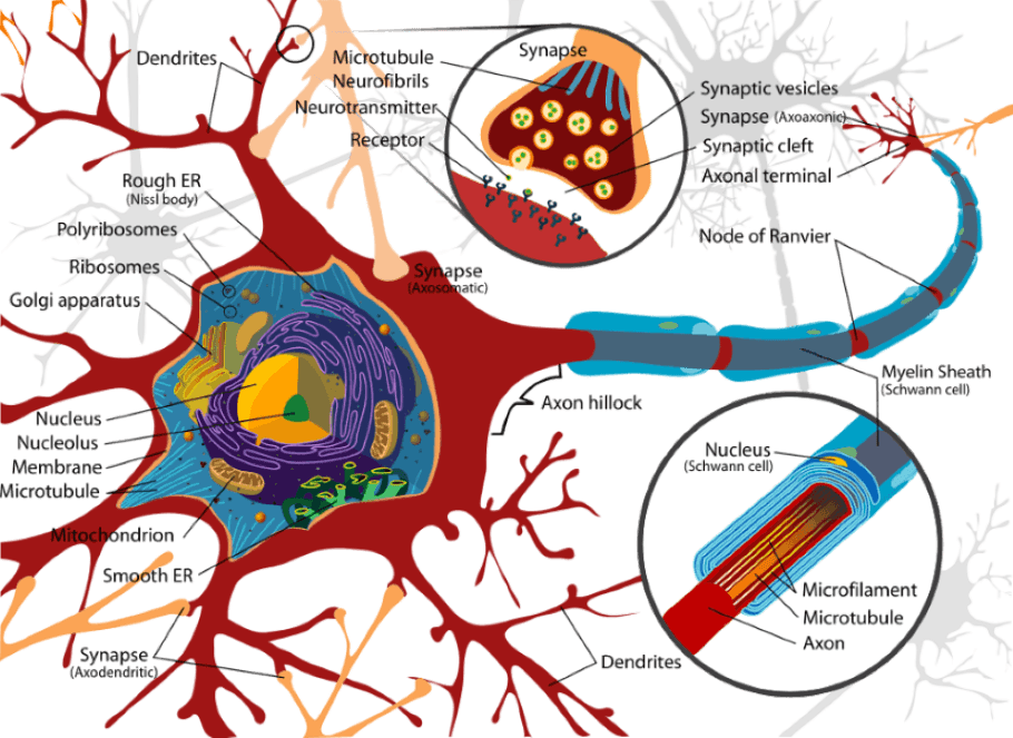
Neurotransmitters
Chemicals that are used to relay, amplify and modulate signals between a neuron and another cell. Neurons expressing certain types of neurotransmitters sometimes form distinct systems, where activation of the system affects large volumes of the brain, called volume transmission. The major neurotransmitter systems are the noradrenaline (norepinephrine) system, the dopamine system, the serotonin system, and the cholinergic system.
Nucleus accumbens
Also known as the accumbens nucleus or as the nucleus accumbens septi, is a collection of neurons within the forebrain. It is thought to play an important role in reward, laughter, pleasure, addiction, fear, and the placebo effect. The medium spiny neuron is the main neuronal cell type in the nucleus accumbens. The neurotransmitter these neurons produce is gamma-aminobutyric acid (GABA), one of the main inhibitory neurotransmitters of the central nervous system. These neurons are also the main projection or output neurons of the nucleus accumbens.
Occipital Lobe
The occipital lobe is the visual processing center of the brain containing most of the anatomical region of the visual cortex. The occipital lobes are the smallest of the four right lobes in the human brain. Located in the rearmost portion of the skull, the occipital lobes are part of the forebrain structure. The lobes rest on the tentorium cerebelli, a dura mater process separating the cerebrum from the cerebellum. They are structurally isolated in their respective cerebral hemispheres by the separation of the cerebral fissure. The lateral occipital sulcus separates the front edge of the occipital several lateral occipital gyri. The occipital aspects along the inside face of each hemisphere are divided by the calcarine sulcus. Above the medial, Y-shaped sulcus lies the cuneus, and the area below the sulcus is the lingual gyrus. Retinal sensors convey stimuli through the optic tracts to the lateral geniculate bodies, where optic radiations continue to the visual cortex. Each visual cortex receives raw sensory information from the outside half of the retina on the same side of the head and from the inside half of the retina on the other side of the head.
Parahippocampal gyrus
The parahippocampal gyrus (or hippocampal gyrus) is a grey matter cortical region of the brain that surrounds the hippocampus. This region plays an important role in memory encoding and retrieval. The anterior part of the gyrus includes the perirhinal and entorhinal cortices. The parahippocampal cortex encompasses the parahippocampal gyrus's posterior portion and the fusiform gyrus's medial portion.
Parietal Lobe
A lobe in the brain. It is positioned above (superior to) the occipital lobe and behind (posterior to) the frontal lobe. The parietal lobe integrates sensory information from different modalities, particularly determining spatial sense and navigation. The parietal lobe plays important roles in integrating sensory information from various body parts, in the knowledge of numbers and their relations, and in manipulating objects. Portions of the parietal lobe are involved with visuospatial processing. Gerstmann's syndrome is associated with a lesion to the dominant (usually left) parietal lobe. Balint's syndrome is associated with bilateral lesions. The hemispatial neglect syndrome is usually associated with large attention deficits in the non-dominant hemisphere.
Pars Compacta
The pars compacta contains neurons which, in humans, are colored black by the pigment neuromelanin that increases with age. This pigmentation is visible as a distinctive black stripe in brain sections and is the origin of the name given to this area. The neurons have particularly long and thick dendrites.
Pars Reticulata
The pars reticulata is a portion of the substantia nigra. They are smaller, thinner than the dopaminergic neurons, and conversely identical and morphologically similar to the pallidal neurons. They connect with the pars compacta dopamine neurons, whose long dendrites plunge deeply into the pars reticulata. The neurons of the pars reticulata and lateralis produce the neurotransmitter gamma-aminobutyric acid (GABA).
Perirhinal cortex
A cortical region in the medial temporal lobe that is made up of Brodmann areas 35 and 36. It receives highly-processed sensory information from all sensory regions and is considered an important memory region. It is bordered caudally by the postrhinal cortex or parahippocampal cortex (homologous regions in rodents and primates, respectively) and ventrally and medially by the entorhinal cortex.
Periaqueductal gray
Midbrain grey matter that is located around the cerebral aqueduct within the midbrain. It plays a role in the descending modulation of pain and defensive behavior. The spinothalamic tract's ascending pain, and temperature fibers also send information to the PAG via the spinomesencephalic tract. The spinomesencephalic tract is so-named because the fibers originate in the spine and terminate in the mesencephalon, another name for the midbrain, which is part of the brain where the PAG resides.
Pineal gland
The pineal gland (also called the pineal body, epiphysis cerebri, or epiphysis) is a small endocrine gland in the vertebrate brain. It produces melatonin, which modulates wake/sleep patterns and photoperiodic (seasonal) functions. It is shaped like a tiny pine cone and is located near the brain's center, between the two hemispheres, tucked in a groove where the two rounded thalamic bodies join.
Pons
A structure located on the brain stem. It is cranial to the medulla oblongata, caudal to the midbrain, and ventral to the cerebellum. The pons relays sensory information between the cerebellum and cerebrum; aid in relaying other messages in the brain; control arousal, and regulate respiration; some also say the pons has a role in dreaming.
Posterior commissure
A rounded band of white fibers crossing the middle line on the dorsal aspect of the upper end of the cerebral aqueduct. It is important in the bilateral pupillary light reflex. Its fibers acquire their medullary sheaths early, but their connections have not been determined. Most of them originate in a nucleus, the nucleus of the posterior commissure (nucleus of Darkschewitsch), which lies in the central gray substance of the upper end of the cerebral aqueduct, in front of the nucleus of the oculomotor nerve.
Postcentral gyrus
The lateral postcentral gyrus is a prominent structure in the parietal lobe of the human brain. Although initially defined to be roughly the same as Brodmann areas 3, 1, and 2, more recent work by Kaas has suggested that for homogeny with other sensory fields, only area 3 should be referred to as "primary somatosensory cortex," as it received the bulk of the thalamocortical projection from the sensory input fields.
Premotor cortex
An area of the motor cortex in the frontal lobe of the brain. It extends 3mm in front of the Primary motor cortex near the Sylvian fissure before narrowing to approximately 1mm near the Medial longitudinal fissure, where it has the prefrontal cortex. It is responsible for sensory guidance of movement and control of the proximal and trunk muscles of the body. It is more or less equivalent to Brodmann area 6.
Primary auditory cortex
The primary auditory cortex is the region of the brain that is responsible for processing of auditory (sound) information.
Primary motor cortex
The primary motor cortex (or M1) works in association with pre-motor areas to plan and execute movements. M1 contains large neurons known as Betz cells, which send long axons down the spinal cord to synapse onto alpha motor neurons which connect to the muscles. Pre-motor areas are involved in planning actions (in concert with the basal ganglia) and refining movements based on sensory input (this requires the cerebellum).
Putamen
The putamen is a structure in the middle of the brain which, together with the caudate nucleus, forms the dorsal striatum. The putamen is a portion of the basal ganglia that forms the outermost part of the lenticular nucleus. The motor and somatosensory cortices, the intralaminar nuclei of the thalamus, and the substantia nigra project to the putamen. The putamen projects to the cortex's premotor and supplementary motor areas via the globus pallidus and thalamus.
Receptors
In biochemistry, a receptor is a protein molecule embedded in either the plasma membrane or cytoplasm of a cell, to which a mobile signaling (or "signal") molecule may attach. In the brain, they sense the environment (chemicals, light, sound, touch) and encode this information into electrochemical messages transmitted by sensory neurons.
Red Nucleus
The red nucleus is a structure in the rostral midbrain involved in motor coordination. It comprises a caudal magnocellular and a rostral parvocellular part. The red nucleus mainly controls the shoulder and upper arm muscles in humans. Still, it has some control over the lower arm and hand as well.
Reticular formation
The reticular formation is a part of the brain that is involved in actions such as waking/sleeping cycle and lying down. It is essential for governing some of the basic functions of higher organisms and is one of the oldest portions of the brain. The reticular formation is an important regulator in the autonomic nervous system for respiration rate, heart rate, and gastrointestinal activity. It also plays an important role in the modulation of sleep, consciousness, and pain.
Spinal cord
The spinal cord is the main pathway for information connecting the brain and peripheral nervous system. The spinal cord is a long, thin, tubular bundle of nerves that extends the central nervous system from the brain and is enclosed in and protected by the bony vertebral column. The main function of the spinal cord is the transmission of neural inputs between the periphery and the brain.
Striatum
The striatum is a subcortical (i.e., inside rather than on the outside) part of the telencephalon. It is the major input station of the basal ganglia system. Anatomically, the striatum is the caudate nucleus and the putamen. The dorsal striatum forms a continuous and large mass, topographically separated by the internal capsule into the caudate nucleus medially, the putamen laterally, and the fundus below, linking the two preceding ventrally, but a single entity. The striatum is homogeneous in terms of neuronal components. It is built up of 4 neuronal types.
Stria medullaris
Also known as stria medullaris thalami, is a fiber bundle containing efferent fibers from the septal nuclei, lateral preoptic-hypothalamic region, and anterior thalamic nuclei to the habenula. It forms a horizontal ridge on the medial surface of the thalamus.
Substantia Nigra
The substantia nigra or locus niger is a heterogeneous portion of the midbrain, separating the pes (foot) from the tegmentum (covering), and an accessory to the basal ganglia system. It consists of two strongly contrasted ensembles, the pars compacta and adjacent dopaminergic groups. Another ensemble made up of the pars reticulate and the pars lateralis.
Sulci
Singular, sulcus, are small grooves, trenches, or furrows, especially fissures of the brain.
Supraoptic nucleus
The supraoptic nucleus (SON) is a nucleus of magnocellular neurosecretory cells in the hypothalamus of the mammalian brain. The nucleus is situated at the base of the brain, adjacent to the optic chiasm, and in humans, it contains about 3,000 neurons.
Superior colliculus
The superior colliculus is a paired structure that is part of the brain's tectal area. Superior Colliculus neurons respond to visual, auditory, and somatosensory stimuli. The two superior colliculi sit below the thalamus and surround the pineal gland in the mesencephalon of vertebrate brains. It comprises the caudal aspect of the midbrain, posterior to the periaqueductal gray and immediately superior to the inferior colliculus. The inferior and superior colliculi are known collectively as the corpora quadrigemina. In humans, the superior colliculus (SC) is involved in the generation of saccadic (fast) eye movements and eye-head coordination.
Synapses
Chemical synapses allow neurons to form interconnected circuits within the central nervous system. They are thus crucial to the biological computations that underlie perception and thought. They provide the means through which the nervous system connects to and controls the other body systems; for example, the specialized synapse between a motor neuron and a muscle cell is called a neuromuscular junction. The adult human brain has been estimated to contain 1014 to 5 Times 1014 (100-500 trillion) synapses.
Tegmentum
The tegmentum is a general area within the brainstem. It is located between the ventricular system and distinctive basal or ventral structures at each level. It forms the floor of the midbrain. It is a multisynaptic network of neurons involved in many unconscious homeostatic and reflexive pathways.
Temporal lobe
Parts of the cerebrum that are involved in speech, memory, and hearing. They lie at the sides of the brain, beneath the lateral or Sylvian fissure. The temporal lobe is involved in auditory processing and is home to the primary auditory cortex. It is also heavily involved in semantics, both in speech and vision. The temporal lobe contains the hippocampus and is therefore involved in memory formation as well.
Tentorium cerebelli
The tentorium cerebelli or cerebellar tentorium is an extension of the dura mater that separates the cerebellum from the inferior portion of the occipital lobes.
Thalamus
A pair and symmetric part of the brain. It constitutes the main part of the diencephalon. The thalamus is generally believed to act as a translator for which various "prethalamic" inputs are processed into a form readable by the cerebral cortex. The thalamus is believed to both process and relay sensory information selectively to various parts of the cerebral cortex, as one thalamic point may reach one or several regions in the cortex. The thalamus also plays an important role in regulating states of sleep and wakefulness. Thalamic nuclei have strong reciprocal connections with the cerebral cortex, forming thalamic-cortical-thalamic circuits that are believed to be involved with consciousness. The thalamus plays a major role in regulating arousal, awareness, and activity. Damage to the thalamus can lead to permanent coma.
Upper motor neuron
Upper motor neurons are motor neurons that originate in the motor region of the cerebral cortex or the brain stems and carry motor information down to the final common pathway, that is, any motor neurons that are not directly responsible for stimulating the target muscle. The main effector neurons for voluntary movement lie within layer V of the primary motor cortex, called Betz cells. The cell bodies of these neurons are some of the largest in the brain, approaching nearly 100um in diameter. These neurons connect the brain to the appropriate level in the spinal cord, from which point nerve signals continue to the muscles using the lower motor neurons. The neurotransmitter glutamate transmits the nerve impulses from upper to lower motor neurons, where glutamatergic receptors detect it.
Ventricles
The ventricular system is a set of structures in the brain, continuous with the central canal of the spinal cord. There are four cerebral ventricles: the paired lateral ventricles and midline, and the third and fourth ventricles. The two lateral ventricles within the cerebrum are relatively large and C-shaped, roughly wrapping around the dorsal aspects of the basal ganglia. In the lateral ventricles of the embryo, the successive generation of neurons gives rise to the 6-layered structure of the neocortex, constructed from the inside out during development. Each lateral ventricle extends into the frontal, temporal, and occipital lobes via the frontal (anterior), temporal (inferior), and occipital (posterior) horns, respectively. The lateral ventricles communicate centrally within the diencephalon via the interventricular foramina with the third ventricle. The third ventricle communicates via the cerebral aqueduct, located within the midbrain, with the fourth ventricle within the hindbrain. The three foramina to the subarachnoid space are found here, permitting cerebrospinal fluid in the ventricles to surround the brainstem, cerebellum, and cerebral cortex. The fourth ventricle is also continuous with the central canal, allowing CSF to bathe the inside surface of the spinal cord as well.
Visual cortex
The term visual cortex refers to the primary visual cortex (also known as striate cortex or V1) and extrastriate visual cortical areas such as V2, V3, V4, and V5. The primary visual cortex is anatomically equivalent to Brodmann area 17, or BA17. The primary visual cortex is the best-studied visual area in the brain. In all mammals studied, it is located in the posterior pole of the occipital cortex (the occipital cortex is responsible for processing visual stimuli). It is the simplest, earliest cortical visual area. It is highly specialized for processing information about static and moving objects and is excellent in pattern recognition.
Wernicke's Area
Part of the human cerebrum that forms part of the cortex on the posterior section of the superior temporal gyrus, encircling the auditory cortex, on the Sylvian fissure (part of the brain where the temporal lobe and parietal lobe meet). For most people, it is located in the left hemisphere, as the left hemisphere is specialized for language skills. Occlusion of the middle cerebral artery in a stroke can affect the proper functioning of this area.
White Matter
One of the three main solid components of the central nervous system. White matter comprises bundles of myelinated nerve cell processes (or axons), which connect the brain's various gray matter areas (the locations of nerve cell bodies) and carry nerve impulses between neurons. Cerebral and spinal white matter do not contain dendrites, which can only be found in grey matter, neural cell bodies, and shorter axons.
Insights, Analysis, and Developments
Editorial Note:It's fascinating to think about how much the brain controls without us even noticing - every step, every memory, every heartbeat tied to this intricate web of cells and signals. Yet, for all its power, the brain's mysteries still feel just out of reach, a reminder of how fragile and resilient we are all at once. This kind of knowledge isn't just for scientists; it's a window into what makes us human, nudging us to appreciate the quiet marvels inside our heads and maybe even push for better ways to protect them as we age or face challenges
. Author Credentials: Ian is the founder and Editor-in-Chief of Disabled World, a leading resource for news and information on disability issues. With a global perspective shaped by years of travel and lived experience, Ian is a committed proponent of the Social Model of Disability-a transformative framework developed by disabled activists in the 1970s that emphasizes dismantling societal barriers rather than focusing solely on individual impairments. His work reflects a deep commitment to disability rights, accessibility, and social inclusion. To learn more about Ian's background, expertise, and accomplishments, visit his full biography.