Guide to Microphthalmia, Anophthalmia and Coloboma
Author: Ian C. Langtree - Writer/Editor for Disabled World (DW)
Published: 26 Mar 2015 - Updated: 10 Oct 2025
Publication Type: Informative
Table of Contents:
Synopsis - Introduction - Main - Insights, Updates - Related Content
Synopsis: This information provides a comprehensive overview of microphthalmia, anophthalmia, and coloboma - congenital eye abnormalities affecting eyeball size and structure present from birth. The resource is authoritative because it synthesizes medical terminology, diagnostic procedures, genetic inheritance patterns, and treatment protocols typically found in clinical ophthalmology references, while making this technical information accessible to general readers.
What makes this particularly useful is its practical coverage of real-world management strategies, including the role of multidisciplinary care teams (pediatricians, geneticists, ocularists, oculoplastic surgeons), the progression of prosthetic eye fittings from infancy through childhood, and detailed explanations of conformers and expanders that help develop eye sockets in affected children. For families navigating these rare conditions - which occur in roughly 1 in 10,000 live births and account for 3-11% of childhood blindness - this material offers clear guidance on what to expect from diagnosis through long-term care.
The information is especially valuable to parents, caregivers, and adults with visual disabilities seeking to understand the medical, genetic, and cosmetic considerations associated with these conditions, providing both clinical context and practical advice about specialist consultations and prosthetic options.
Introduction
Microphthalmia is a form of eye abnormality that arises prior to a person's birth. One or both of a person's eyeballs are smaller than average in this condition. In some people, the eyeball might appear to be missing entirely; however, some remaining eye tissue is usually present.
Main Content
Microphthalmia
Commonly called 'small eye syndrome'. Microphthalmia is a form of eye abnormality that arises prior to a person's birth. One or both of a person's eyeballs are smaller than average in this condition. In some people, the eyeball might appear to be missing entirely; however, some remaining eye tissue is usually present.
Microphthalmia can be either simple or complex.
- Simple microphthalmia refers to an intact eye with shortened total length of the globe
- Complex microphthalmia refers to malformation of various parts of the eye in addition to its small size.
Severe microphthalmia should be distinguished from another condition called, 'Anophthalmia,' in which no eyeball forms at all. The terms, 'anophthalmia,' and, 'severe microphthalmia,' are often times used interchangeably. Microphthalmia may or may not result in notable loss of vision in an affected person.
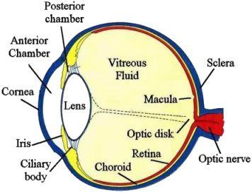
People who experience microphthalmia might also experience a condition called, 'coloboma.' Colobomas are missing pieces of tissue in structures that form a person's eye. They may appear as, 'gaps,' or, 'notches,' in the:
- Colored part of a person's eye called the iris
- Blood vessel layer underneath the retina called the choroid
- Optic nerves, which carry information from the eyes to the brain
- Retina, which is light-sensitive tissue lining the back of the eye
People with microphthalmia may also experience additional eye abnormalities, to include a narrowed opening of the eye or, 'narrowed palpebral fissure,' and clouding of the lens of the person's eye or, 'cataract.' In addition, those affected may have an abnormality referred to as, 'microcornea,' in which the clear front covering of the eye is both small and curved more than average. Between one-third and one-half of people affected have microphthalmia as a part of a syndrome that affects other tissues and organs in their bodies. These forms of the condition are described as being, 'syndromic.' When microphthalmia occurs by itself it is described as being, 'non-syndromic,' or, 'isolated.' These are very rare conditions, occurring in 1 of 10,000 live births. They account for 3 to 11% of blindness in children. About 2/3 of these conditions are considered to be genetic.
Anophthalmia
The term used when there is a total lack of an eye, indicating some outside interruption during intrauterine foetal development very early after conception. Anophthalmia may be monocular (affecting one eye), or bilateral (both eyes). Obviously there is no sight and therefore no treatment can be offered. Cosmetically, however, sockets can be measured, molds taken and artificial eyes (matched with parental or sibling eye coloring) fitted.
Coloboma
A hole in one of the structures of the eye, such as the iris, retina, choroid, or optic disc. The hole is present from birth and can be caused when a gap called the choroid fissure, which is present during early stages of prenatal development, fails to close up completely before a child is born.
Colobomas might be present in one or both of a person's eyes. Depending upon their location and size, they may affect a person's vision.
Genes Related to Microphthalmia
Microphthalmia might be caused by changes in a number of genes involved in the early development of a person's eye; the majority of these genes have yet to be identified. The condition may also result from a chromosomal abnormality that affects one or more genes. The majority of genetic changes associated with isolated microphthalmia have been identified in only a small number of people affected.
It is important to note that microphthalmia may also be caused by environmental factors that affect a person's early development such as:
- Drugs
- Toxins
- X-rays
- Viruses
- Radiation
- Pesticides
- Chemicals
- Infections such as Rubella
- A shortage of vitamins during pregnancy
- Exposure to substances that cause birth defects
Researchers have stated their research is not conclusive. At times, the cause of microphthalmia in a person cannot be determined.
Inheritance of Microphthalmia
Isolated microphthalmia is sometimes inherited in an autosomal recessive pattern, meaning both copies of the gene in each cell have mutations. The parents of people with an autosomal recessive condition each carry one copy of the mutated gene, yet they usually do not show signs and symptoms of the condition itself. In some instances, parents of those affected have less-severe eye conditions.
When microphthalmia occurs as a feature of a genetic syndrome or chromosomal abnormality it might, 'cluster,' in families in accordance with the inheritance pattern for the condition. Microphthalmia may be autosomal recessive or involve other patterns. Often times, microphthalmia is not inherited and there is only one person in a family who is affected.
Diagnosing Microphthalmia or Anophthalmia
A diagnosis of anophthalmia is usually made by a pediatric ophthalmologist or pediatrician during an external examination of a person's ocular structures soon after they are born. The pediatrician opens the person's eyelids to check for the presence of eye tissue. If unilateral, a complete dilated eye examination should be performed on the person's other eye to find out if there are any other eye malformations that may lead to a reduction of the person's vision. The first test to be performed is often times an ultrasound of the person's orbits. Commonly, an MRI of the orbits is needed to make an accurate diagnosis.
Many health care providers should be involved in the care of an infant who has either microphthalmia or anophthalmia. The health care providers assist the infant and their family members to meet challenges related to these conditions to live as healthy a life as they can. The team should include a/an:
- Ocularist
- Geneticist
- Pediatrician
- Genetic counselor
- Oculoplastic surgeon
- Other specialists such as a neurologist or an endocrinologist
A geneticist, who is a doctor with expertise in genetic conditions, as well as a genetic counselor who specialized in support and information sharing, can help a family to coordinate care and early intervention for an affected child. These health care professionals also offer ongoing support and assistance with finding resources in the person's area. The person's genetics team undertakes a detailed evaluation which includes pregnancy and medical history to attempt to identify the cause off microphthalmia or anophthalmia.
While genetic testing might help to achieve a diagnosis, an average chromosomal test does not rule out a genetic cause for microphthalmia or anophthalmia. A person's chromosomes to not show submicroscopic gene changes. As more is being discovered about eye development genes, more genes that cause microphthalmia or anophthalmia are being identified. The genes would not show up in a chromosome study. Considering the quick pace of changes in genetics, an affected child should be seen again by a geneticist in 2-3 years when new information might assist with achieving a diagnosis. The geneticist may also help discuss the potential for other family members to be affected as they are born.
Treatment of Microphthalmia and Anophthalmia
Can microphthalmia and anophthalmia be treated? There is no treatment at this time for severe microphthalmia or anophthalmia that will restore a person's vision or create a new eye for them. People with some less-severe forms of microphthalmia might benefit from medical or surgical treatments. In nearly every instance, improvements to a person's appearance are a possibility.
Children may be fitted for a prosthetic eye for cosmetic purposes and to promote the growth of their eye socket. A newborn with microphthalmia or anophthalmia will need to visit a number of eye care professionals, to include ones who specialize in:
- Pediatrics
- Ophthalmic genetics
- Vitreoretinal disease
- Prosthetic devices for the eye
- Orbital and oculoplastic surgery
Each specialist may provide information and potential forms of treatment, resulting in the best care for both the child and their family members. A specialist in prosthetic diseases for the eye has the ability to make, 'conformers,' which are plastic structures that help support the person's face while encouraging their eye socket to grow. As the person's face continues to develop, new conformers will be required. A child with anophthalmia might also need to use, 'expanders,' along with conformers to further enlarge their eye socket. Once the person's face is fully-developed, prosthetic eyes can be made and placed.
Conformers and Prosthetic Eyes
A painted prosthesis that appears like an average eye is usually fitted between the ages of 1 and 2. Until that time, clear conformers are used. When the conformers are in place, the person's eye socket will appear black. These conformers are not painted to appear like an average eye because they are changed too often.
Every few weeks, a child will progress to a larger size conformer until they reach around two years of age. If a child needs to wear conformers after the age of two, the conformers will be painted like a regular prosthesis, providing the appearance of an average yet smaller eye. The average child with microphthalmia or anophthalmia will need 3-4 new painted prostheses prior to the age of ten.
Children with microphthalmia might have some residual vision. In these instances the person's, 'good eye,' may be patched to strengthen vision in that eye. A prosthesis can be made to cap the eye with microphthalmia in order to assist with cosmetic appearance while preserving the person's remaining sight.
Insights, Analysis, and Developments
Editorial Note: While medical science cannot yet restore vision in cases of severe microphthalmia or anophthalmia, the advances in prosthetic technology and orbital development techniques have transformed quality of life for affected individuals. The multidisciplinary approach outlined here - combining genetics, pediatrics, and specialized ophthalmology - represents a significant shift from earlier decades when cosmetic concerns took precedence over comprehensive care. As researchers continue identifying the genes responsible for these conditions, families gain not only better diagnostic clarity but also more accurate information about recurrence risks and potential interventions. The distinction between syndromic and isolated forms proves particularly crucial for medical management, as roughly one-third to one-half of cases involve additional organ systems requiring coordinated specialist care beyond ophthalmology alone. Author Credentials: Ian is the founder and Editor-in-Chief of Disabled World, a leading resource for news and information on disability issues. With a global perspective shaped by years of travel and lived experience, Ian is a committed proponent of the Social Model of Disability-a transformative framework developed by disabled activists in the 1970s that emphasizes dismantling societal barriers rather than focusing solely on individual impairments. His work reflects a deep commitment to disability rights, accessibility, and social inclusion. To learn more about Ian's background, expertise, and accomplishments, visit his full biography.