Flexible 2D Artificial Retina Could Restore Lost Vision
Author: American Chemical Society (ACS)
Published: 25 Aug 2018 - Updated: 10 Oct 2025
Publication Details: Peer-Reviewed | Research, Study, Analysis
Table of Contents:
Synopsis - Overview - Insights, Updates - Related Content
Synopsis
This peer-reviewed research from the American Chemical Society describes a breakthrough in developing the world's first ultrathin artificial retina using 2D materials like graphene and molybdenum disulfide. Scientists at the University of Texas at Austin and Seoul National University created a flexible, high-density curved sensor array that can conform to the natural shape of the retina - addressing critical limitations of current silicon-based implants, which are rigid, flat, and prone to causing blurry vision or tissue damage. The device successfully absorbed light and transmitted signals through external circuitry in laboratory and animal studies, demonstrating biocompatibility and functional promise.
This work holds particular significance for people with macular degeneration, diabetic retinopathy, and retinitis pigmentosa - conditions that currently have no cure but affect millions worldwide. The research is authoritative because it comes from leading research institutions, was presented at the ACS National Meeting, and has undergone peer review, ensuring scientific rigor. Beyond vision restoration, the technology shows potential for monitoring heart arrhythmias and brain activity, making it relevant not only to those with vision loss but also to people managing cardiac conditions and neurological disorders.*
Overview
Scientists report they have successfully developed and tested the world's first ultrathin artificial retina that could vastly improve on existing implantable visualization technology for the blind. The flexible device, based on very thin 2D materials, could someday restore sight to the millions of people with retinal diseases. And with a few modifications, the device could be used to track heart and brain activity.
The researchers presented their work at the 256th National Meeting & Exposition of the American Chemical Society (ACS). ACS, the world's largest scientific society. It featured more than 10,000 presentations on a wide range of science topics.
"This is the first demonstration that you can use few-layer graphene and molybdenum disulfide to successfully fabricate an artificial retina," Nanshu Lu, Ph.D., says. "Although this research is still in its infancy, it is a very exciting starting point for the use of these materials to restore vision," she says, adding that this device could also be implanted elsewhere in the body to monitor heart and brain activities.
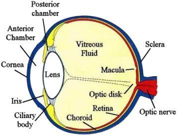
The retina, located at the back of the eye, contains specialized photoreceptor cells called rods and cones that convert incoming light into nerve signals. These impulses travel into the brain via the optic nerve where they are decoded into visual images.
Diseases such as macular degeneration, diabetic retinopathy and retinitis pigmentosa can damage or destroy retinal tissue, leading to vision loss or complete blindness. There is no cure for many of these diseases, but silicon-based retinal implants have restored a modicum of vision to some individuals.
However, Lu says these devices are rigid, flat and fragile, making it hard for them to replicate the natural curvature of the retina. As a result, silicon-based retinal implants often produce blurry or distorted images and can cause long-term strain or damage to surrounding eye tissue, including the optic nerve. Lu, who is at the University of Texas at Austin, and her collaborator Dae-Hyeong Kim, Ph.D., who is at Seoul National University, sought to develop a thinner, more flexible alternative that would better mimic the shape and function of a natural retina.
The researchers used 2D materials, including graphene and molybdenum disulfide, as well as thin layers of gold, alumina and silicon nitrate to create a flexible, high-density and curved sensor array. The device, which resembles the surface of a flattened soccer ball or icosahedron, conforms to the size and shape of a natural retina without mechanically disturbing it.
In laboratory and animal studies, photodetectors on the device readily absorbed light and passed it through a soft external circuit board. The circuit board housed all of the electronics needed to digitally process light, stimulate the retina and acquire signals from the visual cortex. Based on these studies, the researchers determined that this prototype artificial retina is biocompatible and successfully mimics the structural features of the human eye. They say it could be an important step in the quest to develop the next generation of soft bio-electronic retinal prostheses.
Moving ahead, Lu is exploring ways to integrate this technology into mechanically and optically imperceptible electronic tattoos that are laminated on the skin surface to gather real-time health information.
Lu says that the team plans to add transistors to these transparent e-tattoos to help amplify signals from the brain or the heart so they can be more easily monitored and treated. These ultrathin sensors and electrodes can also be implanted on the surface of the heart to detect arrhythmias. Lu says doctors could potentially program them to act like tiny pacemakers, sending electrical impulses through the heart to correct the problem.
Insights, Analysis, and Developments
Editorial Note: What makes this research particularly compelling is not just the technical achievement of creating a flexible retinal prosthesis, but the broader implications for adaptive bioelectronics. The researchers' vision extends beyond the eye itself - imagining a future where imperceptible electronic tattoos monitor our health in real time, or where miniature cardiac devices correct heart rhythms without bulky implants. While still in early development stages, this work represents a fundamental shift in how we think about interfacing technology with human tissue. The use of atomically thin 2D materials solves problems that have plagued medical devices for decades: rigidity, incompatibility with curved biological structures, and tissue damage from mechanical mismatch. As we continue to age as a population and face rising rates of both retinal and cardiovascular diseases, innovations like these may transform from laboratory curiosities into essential medical tools that preserve quality of life.*
Attribution/Source(s): This peer reviewed publication was selected for publishing by the editors of Disabled World (DW) due to its relevance to the disability community. Originally authored by American Chemical Society (ACS) and published on 25 Aug 2018, this content may have been edited for style, clarity, or brevity.
* Editorial additions by Ian C. Langtree.