Brain Skull Connect Implications for Spina Bifida and Chiari Malformation
Author: University of Miami
Published: 2014/11/04 - Updated: 2020/11/29
Contents: Summary - Introduction - Main - Related
Synopsis: Researchers discover network of tissue communication that ensures the brain and spinal cord are matched with the skull and spinal column during embryonic development. The zebrafish, unlike humans and other mammals, can regenerate their spinal cord following injury. Zebrafish also have the ability to regenerate their fins, skin, heart and, in larval stages, brain.
Introduction
Think about the way our bodies are assembled during early development and ask: How do neighboring cells know that they are supposed to become a nerve or a bone cell and how do these tissues find the correct place and alignment? Researchers at the University of Miami (UM) are answering these crucial questions.
Main Digest
In a new study, UM researchers describe the signaling systems that tissues use to communicate with their surrounding neighbors, at the head-trunk region. Their discovery may have important implications for the treatment of congenital defects like Spina Bifida and Chiari malformations.
"Our work describes a network of tissue communication events that ensure that the brain stays in the skull and the spinal cord in the spinal column," said Isaac Skromne, assistant professor of Biology in the UM College of Arts and Sciences and principal investigator of the study.
The findings are published in the November issue of the journal Development in a study entitled "Retinoic acid regulates size, pattern and alignment of tissues at the head-trunk transition."
Study Reports Two Major Findings
- First, it reveals that cells at the head-trunk junction communicate with each other not only to convey information on the type of tissue they will become, but also their location.
- Second, the study finds that signaling the identity and location of the tissues are separate events.
Previous work focused on understanding how tissues acquire their identity, without taking into consideration neighboring tissues.
"That is like knowing the size of each plot of land in a city block, without knowing the addresses," Skromne said. "Now we know the addresses as well, and we show that each plot can take different addresses, potentially changing their relationship to the neighboring plots."
For the study, the researchers analyzed zebrafish embryos, knowing that the findings about the development of this organism would be applicable to other vertebrates, said Keun Lee, first author of the paper and a medical student at the UM Miller School of Medicine. Lee carried out the study when he was an undergraduate student working in Dr. Skromne's lab.
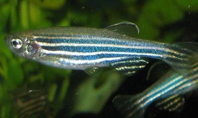
The zebrafish (Danio rerio) is a tropical freshwater fish belonging to the minnow family (Cyprinidae) of the order Cypriniformes. The zebrafish is an important vertebrate model organism in scientific research. It is particularly notable for its regenerative abilities, and has been modified by researchers to produce several transgenic strains.
The zebrafish, unlike humans and other mammals, can regenerate their spinal cord following injury. Zebrafish don't have so much inflammation and the injury is not so severe as in mammals, so we can actually see the pro-regenerative effects that can happen. Zebrafish also have the ability to regenerate their fins, skin, heart and, in larval stages, brain. Zebrafish heart muscle regeneration does not make use of stem cells; instead, mature heart muscle cells regress to a stem cell-like state and re-differentiate. D. rerio is also one of the few fish species to have been sent into space.
"We were hoping to understand the earliest mechanism of organizing nerve and bone-forming tissues in zebrafish embryos, because neuro-skeletal malformation in newborn babies could severely compromise function," Lee said. "Knowing the mechanism of the malformation in the zebrafish model would help develop interventions to prevent those defects in humans."
The findings show that the coordination of brain and nerve tissue at the head-trunk transition in the zebrafish depends on two activities of a signaling molecule called retinoic acid. One activity specifies the size and the other the axial position of the hindbrain territory. In the future, the researchers would like to gain understanding of the type of information these signals carry.
"Now that we have the big picture of how the tissues are coordinated to form the neuro-skeletal system at the head-trunk transition, we would like to know how tissue-specific genes are regulated," Lee said.
The researchers hope that their findings will lead to the development of therapies that target these signaling networks, to prevent abnormalities on the head-trunk junction.
Attribution/Source(s):
This quality-reviewed publication titled Brain Skull Connect Implications for Spina Bifida and Chiari Malformation was chosen for publishing by Disabled World's editors due to its relevance to the disability community. While the content may have been edited for style, clarity, or brevity, it was originally authored by University of Miami and published 2014/11/04 (Edit Update: 2020/11/29). For further details or clarifications, you can contact University of Miami directly at umiami.edu Disabled World does not provide any warranties or endorsements related to this article.
Related Publications
Share This Information To:
𝕏.com Facebook Reddit
Page Information, Citing and Disclaimer
Disabled World is an independent disability community founded in 2004 to provide news and information to people with disabilities, seniors, their family and carers. You can connect with us on social media such as X.com and Facebook.
Cite This Page (APA): University of Miami. (2014, November 4). Brain Skull Connect Implications for Spina Bifida and Chiari Malformation. Disabled World. Retrieved May 19, 2024 from www.disabled-world.com/health/neurology/brain/skull.php
Permalink: <a href="https://www.disabled-world.com/health/neurology/brain/skull.php">Brain Skull Connect Implications for Spina Bifida and Chiari Malformation</a>: Researchers discover network of tissue communication that ensures the brain and spinal cord are matched with the skull and spinal column during embryonic development.
Disabled World provides general information only. Materials presented are never meant to substitute for qualified medical care. Any 3rd party offering or advertising does not constitute an endorsement.