Motor Control Shifts Found After Spinal Cord Injury
Author: KTH, Royal Institute of Technology
Published: 13 Apr 2026
Publication Details: Peer-Reviewed | Observational Study
Table of Contents:
Synopsis - Definition - Overview - Insights, Updates - Related Content
Synopsis
This research, peer-reviewed and published in the Journal of NeuroEngineering and Rehabilitation, examines how incomplete spinal cord injury alters the way the nervous system coordinates muscle movement - even in people who are still able to walk. Using non-invasive skin-surface sensors and high-density electromyography, researchers at KTH Royal Institute of Technology studied motor unit activity in calf muscles during controlled exertion tasks, finding that the injured nervous system struggles to maintain smooth, shared signaling at low effort and overcompensates with rigid, less precise signals at higher effort levels. The findings are particularly relevant for rehabilitation clinicians, physical therapists, and patients with spinal cord injuries, including older adults and those with mobility impairments, as the work may help define new biomarkers for designing better neurorehabilitation strategies.*Topic Definition
- Motor Unit Synchronization
Motor unit synchronization refers to the coordinated firing of groups of motor neurons - each connected to a set of muscle fibers - in response to shared signals from the central nervous system. In a healthy nervous system, this coordination is dynamic and adaptable, allowing muscles to produce smooth, controlled force across a wide range of effort levels. When injury disrupts the spinal cord, these shared neural signals can become either too weak to coordinate muscle activity effectively at low exertion or overly rigid and undifferentiated at higher effort, reducing both precision and flexibility in movement. Studying how synchronization changes after injury provides a measurable window into the functional state of the nervous system and the degree to which it has adapted - or failed to adapt - to the damage it has sustained.
Overview
Study Reveals Unseen Changes In Motor Control After Spinal Cord Injury
Even when people with incomplete spinal cord injuries can walk, everyday functions like standing, balancing or producing steady force may remain difficult. A new study shows why.
Using surface skin electrical sensors, a research team in Sweden identified previously unseen changes in motor coordination that result from incomplete spinal cord injuries (SCI). The study is the first to examine how individual motor units - that is, nerve-to-muscle connections that create movement - work together in people with SCI.
"Our study reveals, at the cellular level, how the central nervous system adapts to the injury to control movement," says Ruoli Wang, associate professor in biomechanics at Promobilia MoveAbility lab, KTH Royal Institute of Technology. She says the researchers' approach was completely non-invasive.
The results were published in the Journal of NeuroEngineering and Rehabilitation.
Adaptation of Motor Unit Synergies In The Synergetic Ankle Plantarflexors In Ambulatory Persons With Incomplete Spinal Cord Injury
The study's lead author, PhD student Zhihao Duan, says the researchers found the nervous system struggles to spread signals smoothly across muscles at low levels of exertion after the injury. And it appears to overcompensate at higher levels of exertion, sending "louder", less refined signals.
A single muscle moves through hundreds of motor units, each turning on and off precisely to create smooth force. Composed of a single motor neuron and its connecting muscle fibers, these motor units respond to shared signals from the nervous system, much like different sections of musicians led by an orchestra conductor. That shared input is what allows them to act in coordinated patterns.
To explore how well these units coordinate under the control of the central nervous system the team examined 25 people (including 10 control participants). They used high density electromyography (HD-EMG) to measure electrical activity in the functionally similar calf muscles - soleus and gastrocnemius - while volunteers pushed lightly or moderately against a force measurement device.
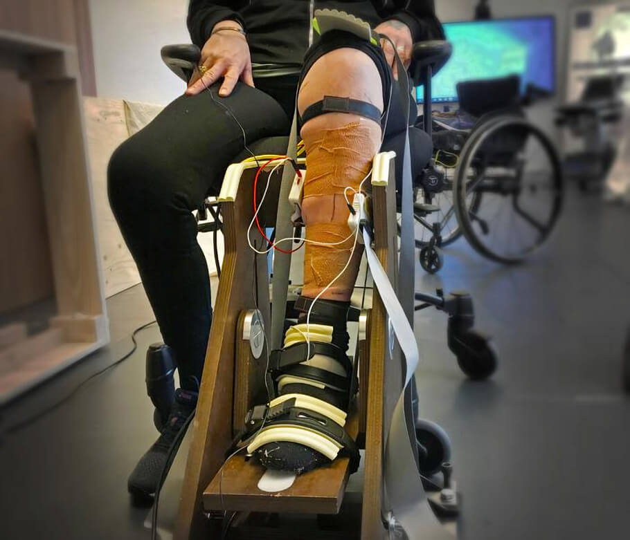
Duan says that at 20 percent effort, fewer of the motor units in the two calf muscles were working in a shared, coordinated way compared with people without injury. Their movements as a result were shaky and unstable.
"They were much less being driven by the same coordinated signal from the nervous system," he says.
At a higher level of effort - 50 percent - the SCI group showed stronger low-frequency synchronization between the two muscles. The body loses flexibility and precision in control of the movement.
"This could be a sign of the nervous system compensating by sending louder, less refined signals," Duan says.
"One interesting finding is that after spinal cord injury the nervous system becomes more rigid and less able to change its approach as the muscles work harder. A healthy nervous system on the other hand is able to adapt its strategy as force demands, to adjust the shared neural drive level," Wang says.
Although the study was limited by a small sample size and challenges in identifying enough motor units per muscle from the skin surface, Wang says the results offer unique insight into how SCI reshapes motor control.
"This finding may open the door to a new rehabilitation biomarker, helping clinicians and researchers design new neurorehabilitation strategies to re-tune the spinal cord control and to restore coordinated neural input," she says.
The study was a collaboration with Aleris Rehab Station and was funded by the Swedish Research Council and Promobilia Foundation.
COI Statement
The authors declare no competing interests.
Insights, Analysis, and Developments
Editorial Note: What makes this study stand out is not just what it found, but how it found it - without a single needle or invasive procedure. The use of high-density electromyography to peer into the nervous system's coordination strategies gives researchers a practical, patient-friendly tool that could realistically be used in clinical settings. The picture that emerges is one of a nervous system doing its best under difficult circumstances, compensating in ways that are ultimately too blunt to restore normal movement. Understanding that distinction - between compensation and true recovery - may prove to be one of the more important conceptual shifts in spinal cord rehabilitation research in recent years.*Attribution/Source(s): This peer reviewed publication was selected for publishing by the editors of Disabled World (DW) due to its relevance to the disability community. Originally authored by KTH, Royal Institute of Technology and published on 13 Apr 2026, this content may have been edited for style, clarity, or brevity.
* Editorial additions by Ian C. Langtree.