Basal Cell Carcinoma on the Finger: A Rare Case Report
Author: Higher Education Press
Published: 26 Jun 2026
Publication Details: Peer-Reviewed | Experimental Study
Table of Contents:
Synopsis - Definition - Introduction - Main - Insights, Updates - Related Content
Synopsis: This article covers a published case report in the journal Skin describing a rare presentation of nodular basal cell carcinoma on the finger of a 63-year-old man, a location where this type of skin cancer seldom occurs. Because digital basal cell carcinoma closely resembles other skin conditions - including melanocytic tumors and squamous cell carcinoma - it is frequently misdiagnosed, making this report a useful clinical reference for anyone seeking to understand atypical skin cancer presentations, early warning signs, and the critical role of biopsy in reaching an accurate diagnosis.*
At a Glance
- 1 - Basal cell carcinoma rarely develops on the fingers due to the low density of sebaceous glands in those areas, making digital BCC an uncommon but serious finding.
- 2 - The lesion in this case was present for three years before diagnosis, underscoring how atypical locations can delay clinical suspicion and proper evaluation.
- 3 - While dermoscopy and reflective confocal microscopy can aid assessment, tissue biopsy remains the definitive method for confirming a basal cell carcinoma diagnosis.
- Topic Definition: Basal Cell Carcinoma
Basal cell carcinoma is a type of non-melanoma skin cancer that originates in the basal cells, which are found in the deepest layer of the epidermis. It is the most commonly diagnosed form of skin cancer worldwide and typically arises on areas of the skin that receive frequent sun exposure, such as the face, scalp, neck, and hands. BCC usually grows slowly and rarely spreads to other parts of the body, but it can cause significant local tissue destruction if left untreated. The condition most often presents as a pearly or waxy bump, a flat scar-like lesion, or a bleeding sore that repeatedly heals and reopens. Treatment typically involves surgical removal, and the prognosis is excellent when the cancer is caught and excised at an early stage.
Introduction
Basal cell carcinoma is the most prevalent form of skin cancer, typically developing on sun-exposed areas like the face and neck. However, BCC affecting the fingers-known as digital BCC-is relatively uncommon, primarily due to the scarcity of sebaceous glands in these regions, which are linked to BCC pathogenesis.
Main Content
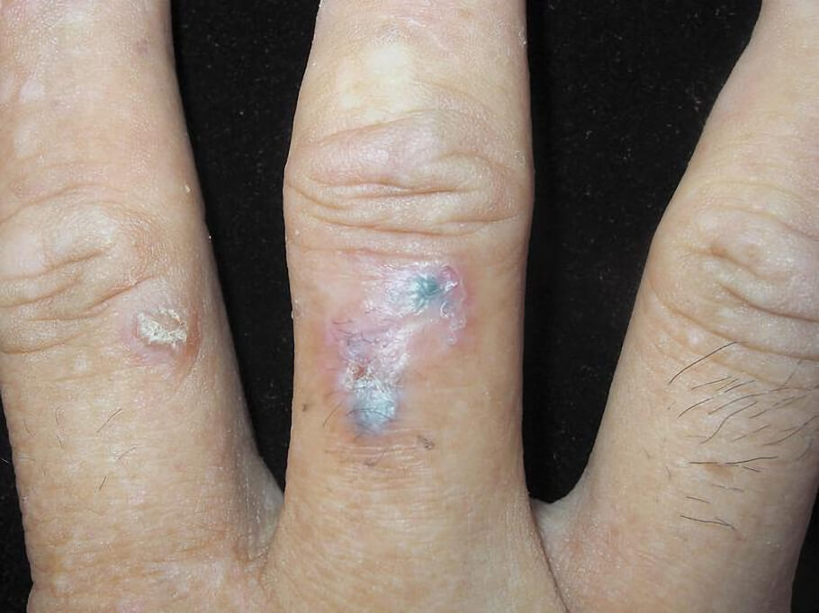
A new case report published in Skin, details a 63-year-old man who sought care for a painful, progressively enlarging lesion on his right middle finger that had been present for three years. Clinical examination showed a 12-mm red plaque topped with two 4-mm bluish-black, firm nodules and surface crusting.
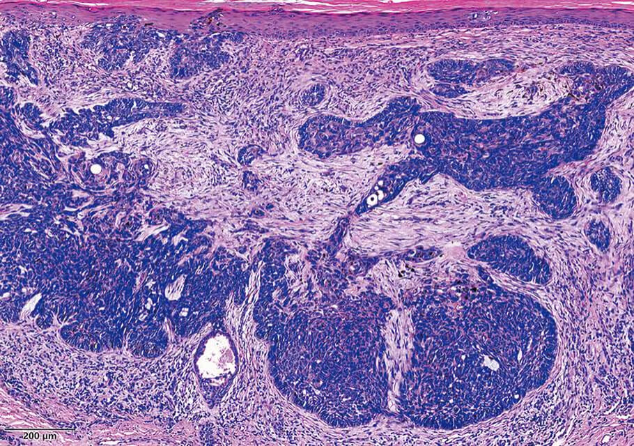
The lesion was surgically excised, and histopathological analysis confirmed a diagnosis of nodular basal cell carcinoma. Microscopic findings revealed tumor nodules composed of basaloid cells with characteristic peripheral palisading and melanin granules within the tumor nests.
The authors note that digital BCC is often misdiagnosed because its appearance mimics other conditions, including melanocytic tumors, squamous cell carcinoma, Bowen disease, and Merkel cell carcinoma. While noninvasive imaging tools such as dermoscopy and reflective confocal microscopy can assist in differential diagnosis, biopsy remains the definitive gold standard.
This case highlights the importance of maintaining clinical suspicion for BCC even in atypical locations. Early recognition and complete surgical excision are essential to prevent local progression and potential complications.
The work titled "Digital Basal Cell Carcinoma" was published in Skin (June 8, 2026).
Insights, Analysis, and Developments
Editorial Note: Digital basal cell carcinoma may be uncommon, but this case is a reminder that skin cancer does not always follow predictable patterns. BCC presenting on the fingers can look deceptively similar to a range of other conditions, and that diagnostic uncertainty carries real consequences - delayed treatment in any form of skin cancer raises the risk of local tissue damage and more complex surgical outcomes. For clinicians and patients alike, the takeaway is straightforward: any persistent, unexplained lesion on the hands warrants proper investigation rather than a watchful waiting approach.*Attribution/Source(s): This peer reviewed publication was selected for publishing by the editors of Disabled World (DW) due to its relevance to the disability community. Originally authored by Higher Education Press and published on 26 Jun 2026, this content may have been edited for style, clarity, or brevity.
* Editorial additions by Ian C. Langtree.